Amazing Science
1.1M views | +23 today
Follow
Amazing Science
Amazing science facts - 3D_printing • aging • AI • anthropology • art • astronomy • bigdata • bioinformatics • biology • biotech • chemistry • computers • cosmology • education • environment • evolution • future • genetics • genomics • geosciences • green_energy • language • map • material_science • math • med • medicine • microscopy • nanotech • neuroscience • paleontology • photography • photonics • physics • postings • robotics • science • technology • video
Your new post is loading...
Scooped by Dr. Stefan Gruenwald
Scoop.it!

Fluorescent Proteins and the Creation of a Living Laser Inside Living Cells

Fluorescent Proteins and the Creation of a Living Laser Inside Living Cells | Amazing Science | Scoop.it

A few years back, a pair of researchers at Massachusetts General Hospital made human cells glow by impregnating them with a molecule that's normally found in jellyfish called green fluorescent protein (GFP) and packing them into a resonant cavity that amplified the amount of light each cell produced. Now, according to a new study recently published in the journal Nano Letters, a team of scientists from the University of St Andrews have developed a means of making individual glowing cells also act as their own resonant cavities.


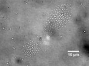
The St. Andrews team accomplished this by coaxing each cell to engulf a tiny plastic bubble (the green dot in the image above) that acts as a resonant cavity. Each bubble is precisely sized and imbued with fluorescent dye. When a laser hits the cell, it excites the dye which bounces around and amplifies inside the bubble, then fluoresces at a different wavelength. Interestingly, the color of the light that the cell emits depends on the size of the bubble. So far, the researchers have gotten cells to produce light at three different wavelengths. And while the team has only been able to get the method to work in petri dishes, they hope to further develop it into a means of tracking specific cells -- say, tumor cells -- for days, even weeks.

No comment yet.
Scooped by Dr. Stefan Gruenwald
Scoop.it!

UCLA develops world’s fastest camera to hunt down cancer in real time

UCLA develops world’s fastest camera to hunt down cancer in real time | Amazing Science | Scoop.it

Engineers at UCLA, led by Bahram Jalali and Dino Di Carlo, have developed a camera that can take 36.7 million frames per second, with a shutter speed of 27 picoseconds. By far the fastest and most sensitive camera in the world — it is some 100 times faster than existing optical microscopes, and it has a false-positive rate of just one in a million — it is hoped, among other applications, that the device will massively improve our ability to diagnose early-stage and pre-metastatic cancer.

No comment yet.
Scooped by Dr. Stefan Gruenwald
Scoop.it!

New biochip technology uses lasers and electric field whirlpools to separate microbes

New biochip technology uses lasers and electric field whirlpools to separate microbes | Amazing Science | Scoop.it

Researchers have demonstrated a new technology that combines a laser and electric fields to create tiny centrifuge-like whirlpools to separate particles and microbes by size, a potential lab-on-a-chip system for medicine and research.


The theory behind the technology, called rapid electrokinetic patterning - or REP - has been described in technical papers published between 2008 and 2011. Now the researchers have used the method for the first time to collect microscopic bacteria and fungi, saidSteven T. Wereley, a Purdue University professor of mechanical engineering.


The technology could bring innovative sensors and analytical devices for lab-on-a-chip applications, or miniature instruments that perform measurements normally requiring large laboratory equipment. REP is a potential new tool for applications including medical diagnostics; testing food, water and contaminated soil; isolating DNA for gene sequencing; crime-scene forensics; and pharmaceutical manufacturing.


"The new results demonstrate that REP can be used to sort biological particles but also that the technique is a powerful tool for development of a high-performance on-chip bioassay system," Wereley said. 


The technology works by using a highly focused infrared laser to heat fluid in a microchannel containing particles or bacteria. An electric field is applied, combining with the laser's heating action to circulate the fluid in a "microfluidic vortex," whirling mini-maelstroms one-tenth the width of a human hair that work like a centrifuge to isolate specific types of particles based on size.


Here the rapid electrokinetic patterning technique is used to arrange bacteria into a specific pattern. The technique may be used as a tool for nanomanufacturing because it shows promise for the assembly of suspended particles, called colloids. The ability to construct objects with colloids makes it possible to create structures with particular mechanical and thermal characteristics to manufacture electronic devices and tiny mechanical parts.


Particles of different sizes can be isolated by changing the electrical frequency, and the vortex moves wherever the laser is pointed, representing a method for positioning specific types of particles for detection and analysis.

No comment yet.